
The Topic of This Month Vol.18 No.4(No.206)
After IASR started to collect information on detection/isolation of pathogens in 1980, adenovirus type 7, of which reports used to be rare, began to be isolated successively from April 1995. Early in 1996, it became apparent that adenovirus type 7 was isolated from fatal pneumonic cases of children with basal ailment in the heart or lung, showing an aspect of emerging infectious diseases in Japan. Consequently, the Ministry of Health and Welfare issued a warning dated April 8, 1996(see p.104 of IASR, Vol.17, No.5). As a topic in the May 1996 issue, IASR took up the actual state of isolation up to February 1996 (see p.99 of IASR,Vol. 17, No. 5). Reports on isolation continued also after that. Papers on serious cases of adenovirus type 7 infection were presented at academic meetings dealing with infections of children by medical institutes in the whole country. Continuous attention should be paid to the trend of this virus. In this topic, detection/isolation of adenovirus type 7 after 1995 will be reported again to draw attention of those who are concerned.
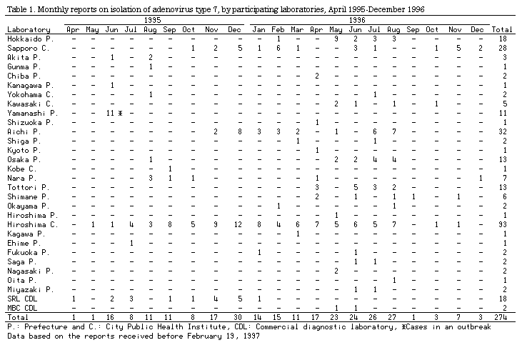
From April 1995 through December 1996, 30 laboratories isolated adenovirus type 7 from 274 cases. Even in the Kyushu district, where there was no report in 1995, it was isolated in 1996 (Table 1).
The specimens from which adenovirus type 7 was isolated comprised 212 nasopharyngeal swabs, 72 stool samples, 30 eye swabs, four urine samples, three lung/bronchus samples, and one thoracic fluid sample (including those from the same cases). The methods for detection were all virus isolation in cultured cells. Eight of these specimens gave also positive results in direct detection of the virus by electron microscopy (EM) and/or antigen detection by ELISA (four cases by EM + ELISA, two cases by EM, and two cases by ELISA). In the serotyping of virus isolates by the neutralization test, attention should be paid to the possible cross reaction arising between type 7 and type 3 or 11 which belong to the same subgenus B (see p.83 of this issue).
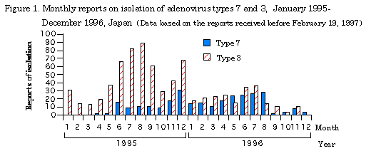
Adenovirus type 7, as does type 3, proliferates in multiple human organs such as the conjunctiva, pharynx, lung, and intestines, developing variety of symptoms. To make comparison with type 3, which has most frequently been isolated among adenoviruses in Japan, the monthly reports on detection of adenovirus types 7 and 3 during 1995-96 are shown in Fig. 1. Type 7 was isolated every month after April 1995, but no distinct seasonal trend was seen. On the other hand, type 3 has been detected mainly in summer every year.
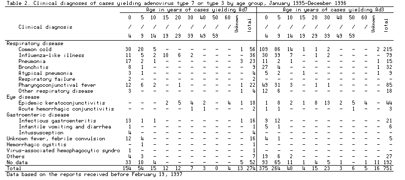
The ages and clinical diagnoses of 274 and 751 cases, from which adenovirus types 7 and 3, respectively, were detected during 1995-96 are shown in Table 2. In 176 cases aged 0 to 14 years with clinical diagnosis recorded, from which type 7 was isolated, respiratory diseases were found in a majority, 137 cases (78%). These cases included 20 pneumonia, two serious cases of respiratory failure and sibling cases (see p. 82 of this issue). Besides, included were 20 cases (11%) of gastrointestinal diseases, 16 cases (9.1%) of unknown fever/febrile convulsion, and one case each of hemorrhagic cystitis and virus-associated hemophagocytic syndrome (VAHS) (see p. 81 of this issue). On the other hand, among 38 cases aged over 15 years, from which type 7 was detected, a large majority were having epidemic keratoconjunctivitis or influenza-like illness (including 11 cases in an outbreak; see p. 101 of IASR, Vol. 17, No. 5 and p. 82 of this issue). Among younger cases, respiratory illness was the main disorder. Among adult cases, eye diseases were observed as was the case with type 3-isolated cases, many type 7-isolated cases had high fever, those with fever higher than 40C accounting for 35% (compared with 27% in type 3-isolated cases).
When pneumonia patients with continuous high fever are examined, it is desired not only to suspect of adenovirus type 7 infection (see p. 4 of IASR, Vol. 18, No. 1), and to take general countermeasures against nosocomial infection, but also to perform rapid diagnosis by ELISA to detect adenovirus antigen in stool samples and pharyngeal swabs (virus isolation is necessary for type identification), to accommodate the patients in separate rooms, and handle patients' stools with care to prevent virus transmission to children with basal ailment in the cardio-pulmonary function or with immunocompromised condition.
 Return to the IASR HomePage
 Return to the IASR HomePage(Japanese)
