![]()
The Topic of This Month Vol.28 No.4(No.326)
In National Epidemiological Surveillance of Infectious Diseases (NESID) complying with the Law Concerning the Prevention of Infectious Diseases and Medical Care for Patients of Infections (the Infectious Diseases Control Law) enacted in April 1999, amebiasis was classified as a category IV notifiable infectious disease, which was then changed to a category V notifiable infectious disease by the partial amendment of the Infectious Diseases Control Law in November 2003. Since April 2006, notification of amebiasis cases requires clarification of the disease type, intestinal and/or extraintestinal amebiasis. On the other hand, reporting asymptomatic cyst carriers is not required (for the criteria of notification, see http://www.mhlw.go.jp/bunya/kenkou/kekkaku-kansenshou11/01-05-01.html).
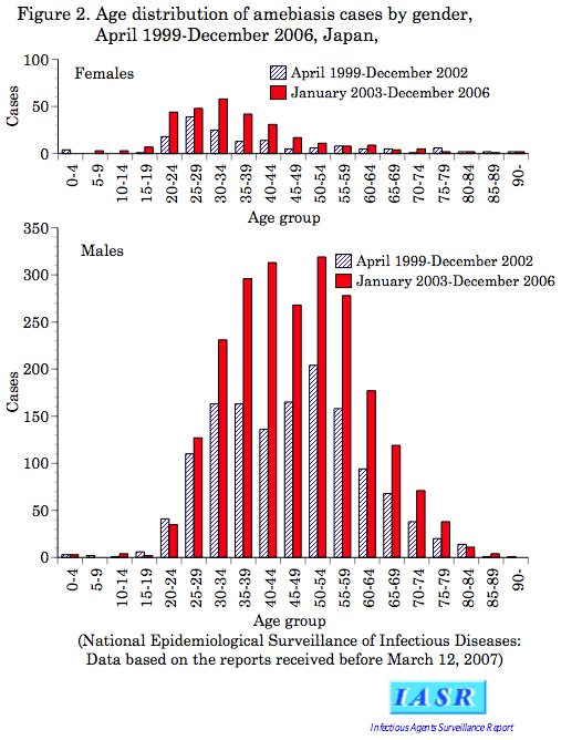
Incidence in Japan based on the NESID: According to WHO, approximately 50 million people, corresponding to about 1% of the total world population, are infected with E. histolytica and it is estimated that each year 40-100 thousand people die of amebiasis. In Japan, annually reported cases during 2000-2002 based on the Infectious Diseases Control Law were 377, 434, and 457 (IASR 24: 79-80, 2003), whereas those during 2003-2006 tended to increase continuously, counting 521, 608, 698 and 747 (Fig. 1). Of the cases reported in 2003-2006, 70% were infected domestically and most of the rest were considered to have been infected in tropical areas, mainly in Southeast Asia. No seasonal or monthly patterns in the occurrence of cases can be seen. This may reflect outbreaks of this disease occurring among male homosexuals (see p. 105 of this issue) and at facilities for the mentally-handicapped (see p. 106 of this issue). As previously, about 90% of cases were males aged 30-60 years, and reports increased 1.7-times compared with those in 1999-2002 (Fig. 2). Female cases were younger than male ones, and the peaks seen at 25-29 years in 1999-2002 were distributed in the wider range of 20-39 years in 2003-2006, and the number of cases increased 1.9-times. Since before, the occurrence of amebiasis among female commercial sex workers (CSW) has been shown (IASR 24: 81, 2003) and investigations in Tokyo have found further spreading of the disease (see p. 108 of this issue). Of the cases diagnosed during 2003-2006, fatal cases at the time of reporting counted at 10, of which nine were males aged 30-80s and the remaining one was a female aged 80s.
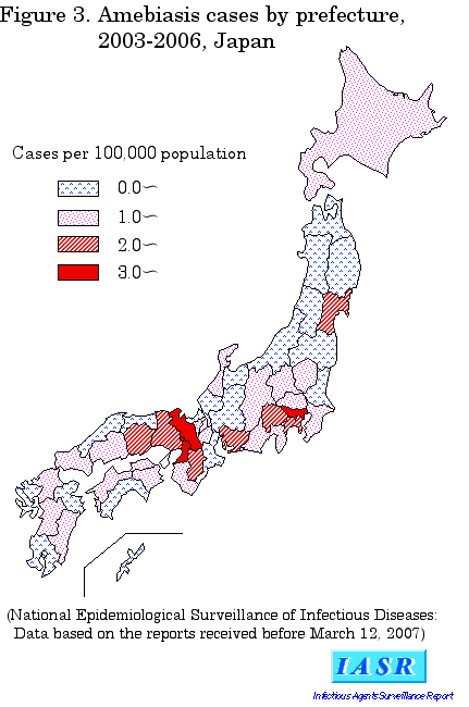
Geographical distribution of reported cases: Among prefectures in which cases were diagnosed and reported during 2003-2006, apparent accumulation of cases was seen in such prefectures having largely populated cities as Tokyo, Kyoto, Osaka, Miyagi, Kanagawa, Yamanashi, Aichi, Hyogo, Nara and Okayama Prefectures (Fig. 3).
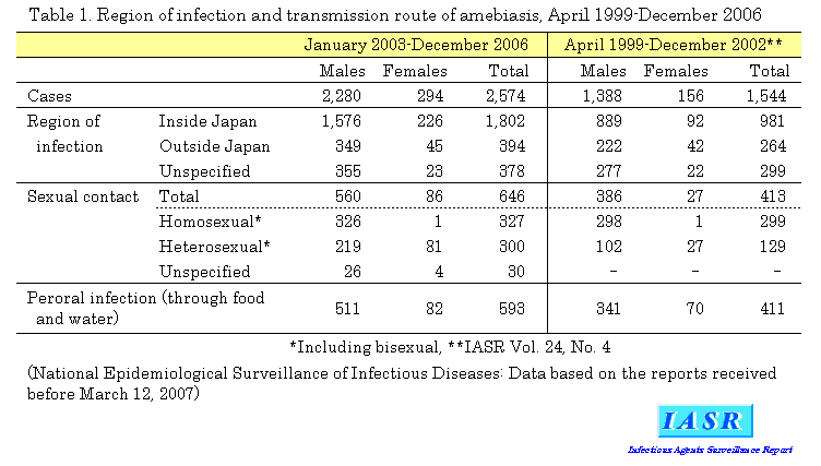
Route of infection: Compared between the terms 1999-2002 (IASR 24: 79-80, 2003) and 2003-2006 (Table 1), domestically-acquired infections (1.8-times) rather than foreign-acquired ones (1.5-times) were on the increase (Fig. 1). Cases due to sexual contact increased 1.6-times and oral infection 1.4-times. It is noteworthy that male cases due to heterosexual contacts increased 2.1-times. Besides, female cases infected by sexual contact increased as largely as 3.2-times, although the actual number of the cases was small.
Diagnosis, types of disease and treatment: For amebae parasitizing human intestines, nonpathogenic E. dispar does not require treatment but differential diagnosis from E. histolytica is important, although morphological differentiation is not possible. Currently, diagnosis can be made by (1) detection of trophozoites or cysts in the stool, colonic mucosa or abscess fluids, (2) detection of specific antigen by ELISA, (3) detection of E. histolytica DNA by PCR (see p. 110 of this issue), (4) detection of serum antibodies, or (5) colonoscopy, ultrasound, or CT scan. The most reliable methods at present to differentiate E. histolytica are (2) and (3), and for (2), E. histolytica II kit (TechLab, USA) is commercially available. We have to be cautious that a serological positivity with the above (4) indicates history of infection but not always the current infection (for detailed method of diagnosis, refer to the manual of infectious agent detection, http://www.nih.go.jp/niid/reference/pathogen-manual-60.pdf#218).
According to the current criteria of notification, diagnosis must be confirmed by showing the presence of E. histolytica with either of the above methods (1)-(5). About half of the reported cases were based on clinical symptoms and serodiagnosis, without detecting the pathogen itself. Detection of specific antigen or specific DNA was performed only in rare cases, and both morphological identification and serological detection were performed only in 20%. Interestingly, diagnosis of amebiasis was obtained after stool occult blood tested positive during health check in 10-odd asymptomatic cases.
As for the disease types reported since April 2006, extraintestinal amebiasis accounted for 19% of all cases; 12% of female and 20% of male cases, thus in a larger proportion of male cases than female ones (see p. 109 of this issue). As pointed out before, intestinal symptoms were rarely seen in extraintestinal amebiasis cases; only 10 to 20% of those cases showed intestinal symptoms as well. Conversely, a few per cent of cases reported as intestinal amebiasis were complicated with extraintestinal symptoms.
Usually, oral administration of metronidazole is the mainstay of treatment of amebiasis and its therapeutic efficacy is high (see p. 105 of this issue). For cyst carriers and cases in the facility with recurrent infections, diloxanide furoate or paromomycin, which is poorly adsorbed from the intestine, is used as well (see p. 106 of this issue).
Control measures in future: Although notification is not required for asymptomatic cyst carriers, diagnosis and treatment of those carriers are important for control measures against the infection. In Japan, outbreaks of amebiasis among male homosexuals and in facilities for the mentally-handicapped have been focused; infection is also expanding to CSW in recent years. Since many cases of amebiasis in our country occur together with such sexually transmitted infections as syphilis, HIV or hepatitis B (see p. 105 and 109 of this issue), control measures against amebiasis should be carried out as part of the comprehensive ones which target overall sexually transmitted infections.
 Return to the IASR HomePage
 Return to the IASR HomePage(Japanese)

 Return to the TopPage