![]()
The Topic of This Month Vol. 32, No. 3 (No. 373)
Trends in infectious gastroenteritis under the National Epidemiological Surveillance of Infectious Diseases: Rotavirus infection is included in “infectious gastroenteritis”, a Category V infectious disease under the Infectious Diseases Control Law. The “infectious gastroenteritis” cases are reported from approximately 3,000 pediatric sentinel clinics nationwide. As “infectious gastroenteritis” is a syndromic category, it is caused by many infectious agents including rotavirus. Prefectural and municipal public health institutes (PHIs) conduct laboratory diagnosis of the infectious gastroenteritis with fecal specimens sent from ca. 10% of the pediatric sentinel clinics and also with specimens collected from outbreak cases.
Every year, the reports of infectious gastroenteritis cases increases sharply during November/December, reaches a peak in January/February, and the decreases from March to May (see http://idsc.nih.go.jp/idwr/kanja/weeklygraph/04gastro.html). The peak of rotavirus is preceded by the peak in November/December of norovirus (see IASR 31: 312-314, 2010 and http://idsc.nih.go.jp/iasr/prompt/graph-ke.html). Among sporadic cases of infectious gastroenteritis during 2005-2010, norovirus, rather than rotavirus, prevailed in all age groups, though rotavirus cases were relatively frequent among young children (Fig. 1).
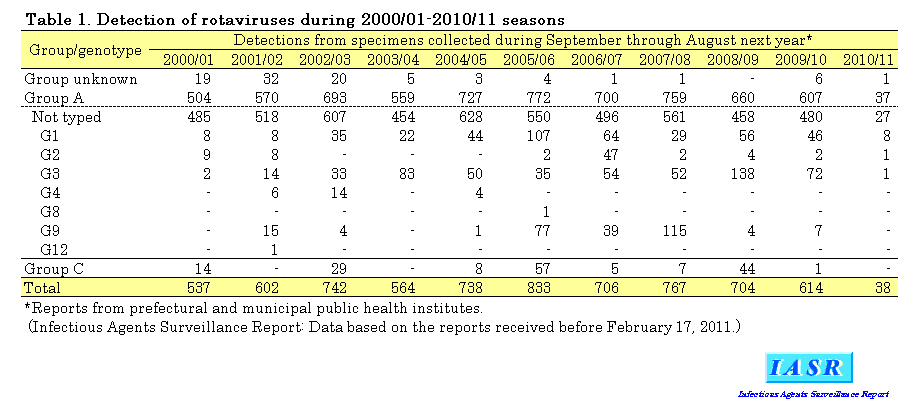
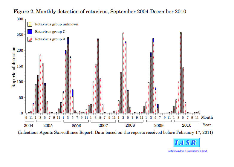
Reports of rotavirus detection from PHIs: Rotavirus belongs to the family Reoviridae , a group of viruses with double-stranded segmented RNA genomes. Rotavirus is classified into groups A-G, among which groups A-C are detected from humans (see p. 63 of this issue). During 2005-2010, 59 PHIs reported group A and 17 PHIs reported group C viruses. Every year 600-800 rotaviruses have been detected by PHIs (Table 1). Among them, group A has been the majority and group C the minority (2-3% of the detections, except in 2005/06 and 2008/09 when relatively higher numbers of group C viruses were reported). No group B rotavirus has been reported in Japan. The peak detection of group A rotaviruses was between in March-April (Fig. 2).
Genotyping of group A rotavirus: Group A rotavirus is further classified according to G genotype which is defined by coat protein VP7 (there are types 1-15, among which types 1-6, 8-12 are known in humans) and P genotype which is defined by VP4 (there are types 1-26, among which types 3-6, 8-11, 14, 19 and 25 are known in humans). The both G and P genotypes are considered to reflect the serotype/antigenicity of the viruses (see p. 64 of this issue).
From 2005 to 2010, 25 PHIs reported 1,053 G genotyped viruses, which were 25% of group A rotaviruses. The most frequent was G1 followed by G9 and G3 in 2005/06, G1 followed by G3 and G2 in 2006/07, and G9 followed by G3 in 2007/08. In 2008/09, the majority was G3 (Table 1). Among prefectures reporting more than 100 cases, i.e., Okayama (464 cases, see p. 71 of this issue), Aichi (177 cases, see p. 72 of this issue) and Niigata (167 cases, see p. 73 of this issue), frequencies of G genotypes fluctuated, in 2007/08, in particular, G9 dominated in all of the three prefectures, suggesting that G9 prevailed in all parts of Japan in that season. In 2006, G8 was reported for the first time in Japan from a patient who had no history of oversees travel.
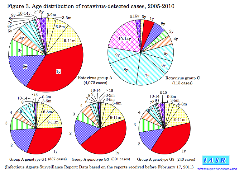
Age distribution of rotavirus-detected cases (Fig. 3): Of the 4,072 cases from which group A rotavirus was detected during 2005-2010, 38% were children one year of age, 20% were those less than one year of age, and 16% were those two years of age, thus children under three years of age occupying the three fourth. Infants aged 6 months or older occupied 80% of 0-year olds from which group A rotavirus was detected. The same age distribution was observed irrespective of genotypes G1, G3 or G9. Of the 115 group C rotavirus-detected cases, 57% were children 5-9 years of age and 20% were those 10-14 years of age.
Encephalitis/encephalopathy cases: Group A rotavirus was detected from feces of 14 cases of encephalitis/ encephalopathy (G3 from 4 cases; G1 from 3 cases; G2 from 1 case; genotype undetermined from 6 cases). Among them, 2 encephalitis cases (IASR 27: 279-280, 2006; ibid. 31: 214, 2010) were positive in the PCR test of their cerebrospinal fluids. In addition, from cerebrospinal fluids of 2 meningitis cases group A rotavirus was detected.
Outbreak incidents: While rotavirus gastroenteritis occurs mainly in children 0-2 year of age, outbreaks in nursery schools, kindergartens, primary schools, hospitals, nursing homes, and welfare facilities were not infrequent (IASR 26: 100-101, 2005; ibid. 26: 339 & 340, 2005; ibid. 27: 153-154, 154-155 & 155, 2006; ibid. 29: 132-134, 2008; ibid. 30: 185-186, 2009 and p. 74 & 75 of this issue). During 2005-2010, group A rotavirus caused 62 outbreaks (9 G1 incidents; 4 G3 incidents; 3 G2 incidents; 1 G9 incident; 45 incidents of G genotype unidentified) and group C rotavirus caused 31 outbreaks. These incidents were all caused by person-to-person transmission except for two food poisoning outbreaks caused by group A rotavirus (IASR 27: 156, 2006). There were seven outbreaks involving more than 50 persons; all outbreaks were in primary schools caused by group C rotavirus (4 incidents from February to May in 2006; one incident each in May 2007, in March 2008 and in March 2009) (IASR 27: 121-122, 2006; ibid. 30: 134-135, 2009).
Prevention and Countermeasures: Rotavirus-infected persons shed as many as 1010 virions per gram stool thus regarded as highly infectious. Proper disposal of diapers, hand washing, and disinfection of contaminated clothing with hypochloride are the basics for prevention of rotavirus infection spread.
Currently two oral attenuated live vaccines are approved abroad; one is G1P[8] monovalent vaccine and the other is pentavalent vaccine containing G1-G4 and P[8] antigens (see p. 68 of this issue). They are used on more than 100 countries world wide. The United States has incorporated the vaccine in the routine immunization for the purpose of reducing the severe cases.
Challenges: Since middle of 1980's, individual medical facilities started to conduct a rapid laboratory diagnosis using simple group A rotavirus antigen detection kits. As a consequence, specimens negative in such rapid tests may be selected and sent to PHIs. It is therefore necessary to investigate the practices in medical facilities to check this possibility.
When vaccines are introduced in Japan, evaluation should be conducted in conjunction. Such evaluations necessitate epidemiological data on rotaviruses before and after the introduction of the vaccine (see p. 68 & 69 of this issue). Such epidemiological data should be consolidated by laboratory data of specimens obtained from gastroenteritis patients, particularly of detection of group C viruses and genotyping of group A viruses, and other genetic characters. In Japan only a few laboratories in NIID, PHIs and universities are conducting sequence analysis of rotavirus. Further improvement of surveillance systems need to be addressed.
 Return to the IASR HomePage
 Return to the IASR HomePage(Japanese)


 Return to the TopPage